定位你的能量小体MitoSpy™ Mitochondrial Probes

免疫荧光(IF)的应用
MitoSpy™ Orange和 MitoSpy™ Red定位于线粒体是基于膜电位,因此MitoSpy™ Orange和 MitoSpy™ Red能很好的反应细胞的健康状态和定位。MitoSpy™Orange CMTMRos和MitoSpy™Red CMXRos含有一个氯甲基团,它能共价与细胞中的半胱氨酸连接,这使得细胞在经过固定破膜后,仍能保持在细胞中。但由于固定破膜过程中洗掉了大部分的试剂,所以这需要使用更高的试剂浓度来获得充分的染色(具体浓度可参考下表)。
而MitoSpy™ Green FM不同于之前的两个,它定位于线粒体不是基于膜电位。它不含有氯甲基,而是含有氟甲基,所以固定破膜之后,大部分的MitoSpy™ Green会被洗掉。所以需要固定破膜的样本,不建议使用该试剂。
|
细胞条件 |
MitoSpy™Orange推荐浓度 |
MitoSpy™Red推荐浓度 |
MitoSpy™ Green推荐浓度 |
|
活细胞 |
50-250nM |
50-250nM |
50-250nM |
|
固定的细胞 |
50-250nM |
50-250nM |
50-250nM |
|
固定破膜后的细胞 |
250-500nM |
250-500nM |
不推荐 |
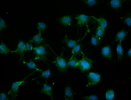
NIH3T3 cells were stained with 100 nM of MitoSpy™ Red CMXRos (red) for 20 minutes at 37°C, fixed with 1% paraformaldehyde (PFA) for ten minutes at room temperature, and permeabilized with 1X True Nuclear™ Perm Buffer for ten minutes at room temperature. Then the cells were stained with Flash Phalloidin™ NIR 647 (green) for 20 minutes at room temperature and counterstained with DAPI (blue). The image was captured with a 60x objective
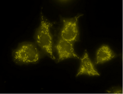
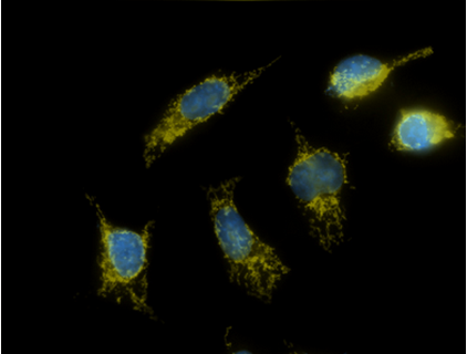
Live staining (50 nM) Fix/Perm staining(500nM)
MitoSpy™ Orange
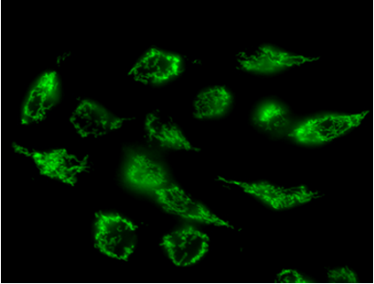
HeLa cells that were stained live with either MitoSpy™ Orange (yellow) or Green (green). Cells that were fixed and permeabilized with 4% PFA and 0.1% Triton X-100 were also stained with DAPI (blue). Photos were taken with a 60x magnification.
流式应用(FC)
MitoSpy™Orange CMTMRos和MitoSpy™Red CMXRos也可作为细胞健康的指标。当线粒体活跃的呼吸时,线粒体膜之间存在潜在的差异,称为膜极化。如果细胞正经历凋亡或死亡,则该探针不会被该细胞的线粒体强烈吸引。如下图所示,MitoSpy™Orange和MitoSpy™Red的阳性细胞群是活细胞和健康的细胞,而Annexin V阳性细胞群是凋亡的早期阶段的细胞。在流式应用中,这两种试剂不应在分析前被固定,因为固定会有大量的试剂流失。如果试剂经过固定后丢失,信号强度的损失会混淆确定表型凋亡的能力。所以 MitoSpy™Orange和MitoSpy™Red的固定仅适用于成像应用中的亚细胞定位。
而对于MitoSpy™ Green,但现在MitoSpy™ Green FM的完整机制还不是很清楚,可能与该试剂的结构和线粒体表面的蛋白之间的同源性相关。由于该试剂的膜电位的独立性,可以用于流式中测量单个细胞的线粒体量。
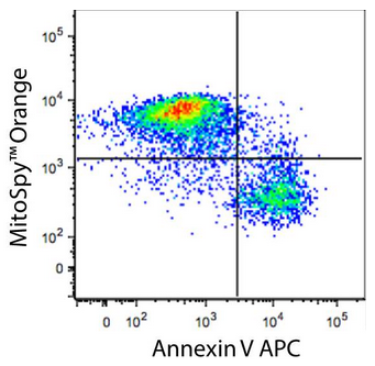
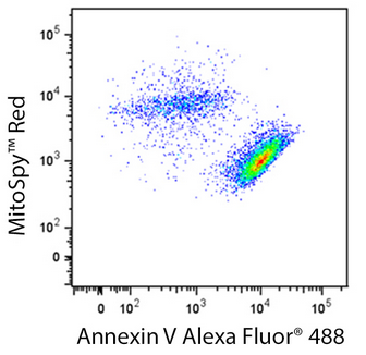
Human T-cell leukemia cell line, Jurkat, was treated for 5 hours with LEAF™ purified anti-CD95 (clone EOS9.1), then stained with an impermeant nucleic acid stain, APC or Alexa Fluor® 488 Annexin V, and either MitoSpy™ Orange or MitoSpy™ Red as indicated. Nucleic acid stain positive events were excluded from analysis.
|
|
|
|
|
|
适合细胞类型 |
适合样本 |
应用 |
固定 |
|||
|
Cat# |
产品名称 |
Ex/Em |
等效通道 |
亚细胞定位 |
活细胞 |
死/固定细胞 |
组织 |
细胞 |
流式 |
显微镜 |
用PFA处理* |
|
424805/ 424806 |
MitoSpy™ Green FM |
488 nm/520 nm |
FITC |
线粒体 |
● |
|
|
● |
● |
● |
|
|
424803/ 424804 |
MitoSpy™ Orange CMTMRos |
551 nm/576 nm |
Alexa Flour®555,PE |
线粒体 |
● |
|
|
● |
● |
● |
● |
|
424801/ 424802 |
MitoSpy™ Red CMXRos |
577 nm/598 nm |
Alexa Flour®594 |
线粒体 |
● |
|
|
● |
● |
● |
● |


